5 July 2020
New Research: Identification of ALK in Thinness

When it comes to obesity research, most geneticists focus on finding gene variants that are associated with increased body weight. In the paper, the researchers did exactly the opposite. They searched for genes that are associated with thinness. And they were very successful and identified a gene called ALK (which stands for Anaplastic lymphoma kinase).
The gene product of ALK is a protein that belongs to the family of growth factor receptors. ALK sits on the cell membrane and when a ligand binds to it, it dimerizes and then activates downstream signaling pathways. ALK is famous for being highly activated in different types of cancer, in particular in lung cancer. ALK activation is due to a chromosomal translocation that results in ALK being fused to another cytosolic protein. The most common fusion partner in ALK-positive lung cancer is the echinoderm microtubule-associated protein like-4 (EML4), a microtubule destabilizing protein. Because EML4 self-associates, the ALK fusion proteins can form homodimers even in the absence of a ligand. This makes ALK constitutively active, promoting tumor formation.
In the new paper, the authors identified a new role for ALK in the brain, where it functions as a thinness gene. They looked for gene variants that are associated with low body weight by comparing an Estonian population of metabolically healthy thin individuals (lowest 6th percentile of the body mass index (BMI) spectrum) with control individuals (which were within the 30–50th percentile for the BMI). They found a marked difference in the frequency if two genetic ALK variants in thin versus normal subjects. This suggests that certain mutations in the ALK gene affect the body weight.
To study the functional role of ALK, the researchers deleted ALK in fruit flies and in mice. This resulted in flies with decreased triglyceride levels and thin mice with marked resistance to obesity. They also found that the decreased body weight in ALK mutant mice is a consequence of increased energy expenditure and elevated lipid breakdown in adipose tissue.
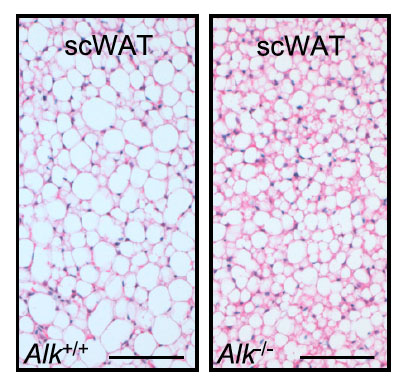
Representative pictures of hematoxylin and eosin stained adipose tissue sections in normal mice (Alk+/+) and mice with deleted ALK (Alk-/-). The results show that ALK deficient mice have smaller fat cells in their adipose tissue.
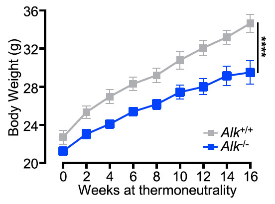
Body weight gain of male normal mice (Alk+/+) and mice with deleted ALK (Alk-/-) over time. The results show that ALK deficient mice gain less weight over time.
The authors then investigated where in the body ALK may function to regulate the body weight. They found that ALK was not expressed in key metabolic tissues, such as the liver, muscle or adipose tissue. However, ALK was expressed in a specific type of nerve cells in the hypothalamus in the brain, suggesting that ALK acts in the brain. They confirmed this by showing that specifically deleting ALK in the hypothalamus, but not in other tissues, caused resistance of mice to high fat diet induced obesity.
The results suggest a mechanism whereby ALK expressing nerve cells in the hypothalamus normally negatively regulate lipid breakdown in adipose tissue. This is likely mediated by inhibiting sympathetic nerve signaling. Indeed, ALK mutant mice as well as thin human individuals were found to have markedly higher norepinephrine/noradrenaline levels in their adipose tissue.
One obvious question is if the reverse true in obese individuals. In other words, do overweight individuals have higher ALK activity. The findings of course also raise the possibility of finding ALK inhibitory drugs to treat obesity, for which the researchers have already registered a patent. This was indeed a really interesting paper!
Original PDF